MICCAI 2018 - Machine Learning in Medical Imaging - doi: 10.1007/978-3-030-00919-9_39
Authors - Soheil Esmaeilzadeh, Dimitrios Ioannis BelivanisKilian, M. Pohl, and Ehsan Adeli
Download the Conference Version Download the arXiv Version Featured on the News
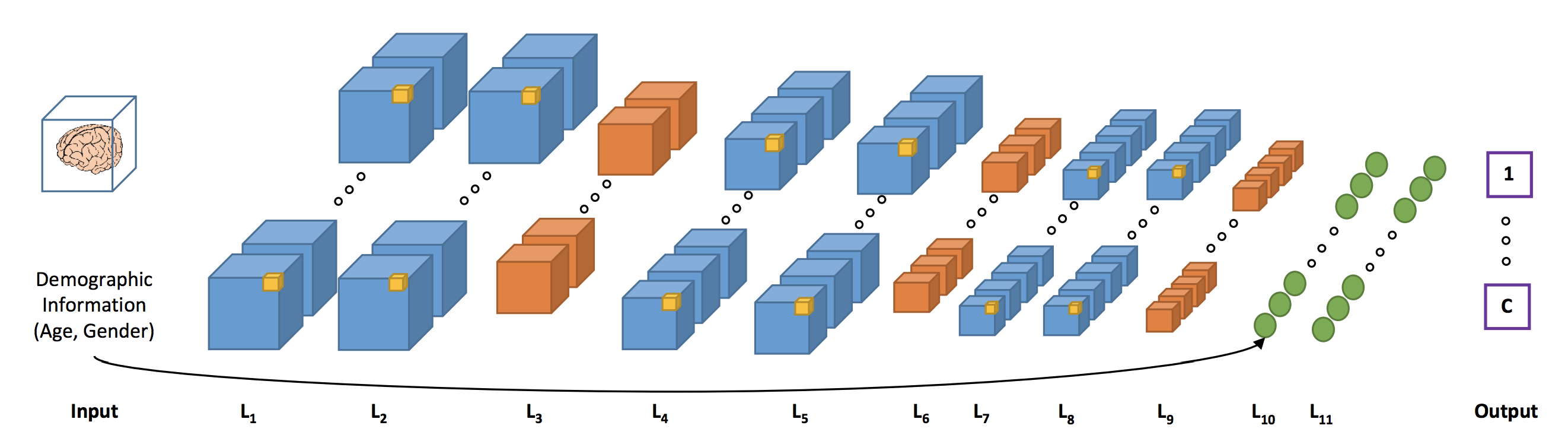
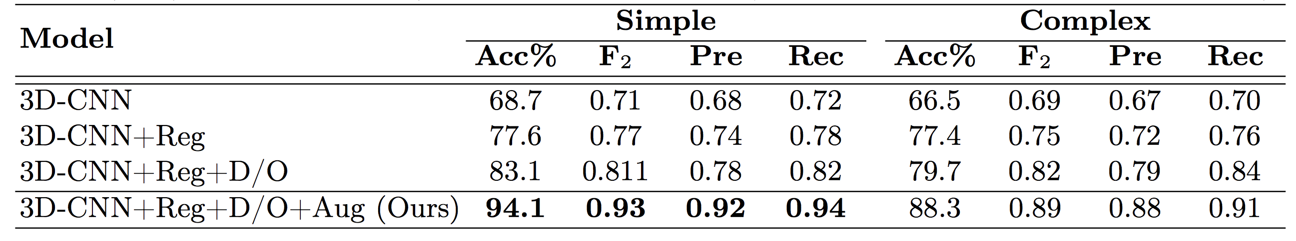
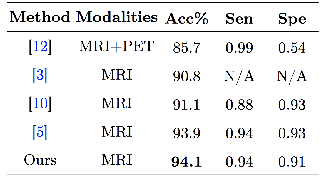
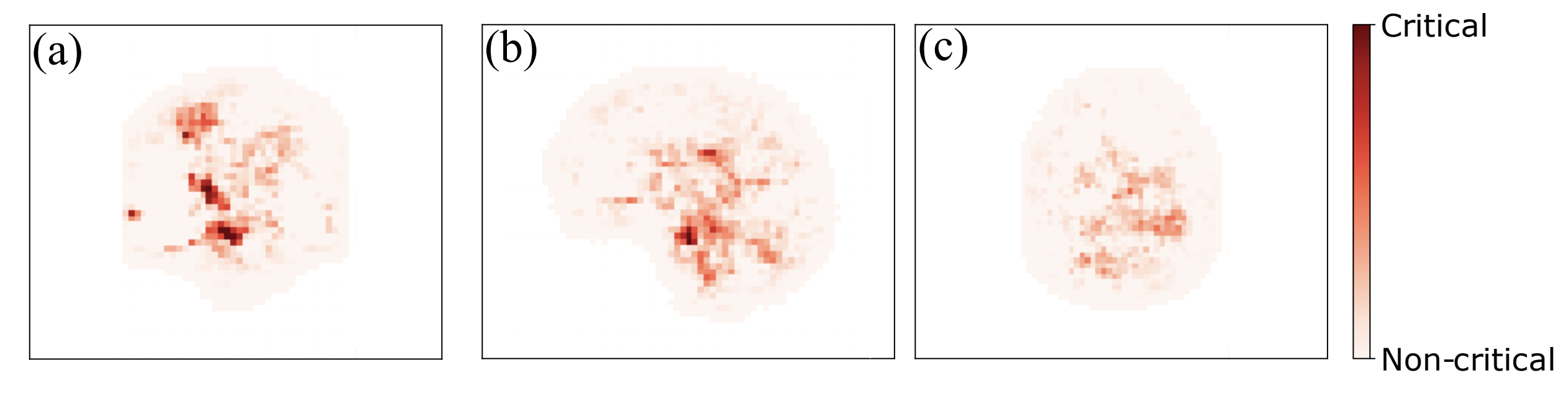
Abstract - As shown in computer vision, the power of deep learning lies in automatically learning relevant and powerful features for any perdition task, which is made possible through end-to-end architectures. However, deep learning approaches applied for classifying medical images do not adhere to this architecture as they rely on several pre- and post-processing steps. This shortcoming can be explained by the relatively small number of available labeled subjects, the high dimensionality of neuroimaging data, and difficulties in interpreting the results of deep learning methods. In this paper, we propose a simple 3D Convolutional Neural Networks and exploit its model parameters to tailor the end-to-end architecture for the diagnosis of Alzheimer’s disease (AD). Our model can diagnose AD with an accuracy of 94.1% on the popular ADNI dataset using only MRI data, which outperforms the previous state-of-the-art. Based on the learned model, we identify the disease biomarkers, the results of which were in accordance with the literature. We further transfer the learned model to diagnose mild cognitive impairment (MCI), the prodromal stage of AD, which yield better results compared to other methods.
Keywords: Alzheimer disease, Brain MR-Image, Deep-learning, Computer vision